Click image to see more details
Product Info Summary
| SKU: | A01478-1 |
|---|---|
| Size: | 100 µg/vial |
| Reactive Species: | Human |
| Host: | Rabbit |
| Application: | IHC |
Customers Who Bought This Also Bought


Product info
Product Name
Anti-S100A12 Antibody Picoband™
View all EN-RAGE/S100A12 Antibodies
SKU/Catalog Number
A01478-1
Size
100 µg/vial
Form
Lyophilized
Description
Boster Bio Anti-S100A12 Antibody Picoband™ catalog # A01478-1. Tested in IHC application. This antibody reacts with Human.
Storage & Handling
At -20°C for one year from date of receipt. After reconstitution, at 4°C for one month. It can also be aliquotted and stored frozen at -20°C for six months. Avoid repeated freezing and thawing.
Cite This Product
Anti-S100A12 Antibody Picoband™ (Boster Biological Technology, Pleasanton CA, USA, Catalog # A01478-1)
Host
Rabbit
Contents
Each vial contains 4 mg Trehalose, 0.9 mg NaCl, 0.2 mg Na2HPO4.
Clonality
Polyclonal
Clone Number
1B9
Isotype
IgG
Immunogen
A synthetic peptide corresponding to a sequence at the C-terminus of human S100A12.
*Blocking peptide can be purchased. Costs vary based on immunogen length. Contact us for pricing.
Cross-reactivity
No cross reactivity with other proteins.
Reactive Species
A01478-1 is reactive to S100A12 in Human
Applications
A01478-1 is guaranteed for IHC Boster Guarantee
Observed Molecular Weight
56,60 kDa
Calculated molecular weight
10.575kDa
Background of EN-RAGE/S100A12
S100 calcium-binding protein A12 (S100A12) is a protein that in humans is encoded by the S100A12 gene. The protein encoded by this gene is a member of the S100 family of proteins containing 2 EF-hand calcium-binding motifs. S100 proteins are localized in the cytoplasm and/or nucleus of a wide range of cells, and involved in the regulation of a number of cellular processes such as cell cycle progression and differentiation. S100 genes include at least 13 members which are located as a cluster on chromosome 1q21. This protein is proposed to be involved in specific calcium-dependent signal transduction pathways and its regulatory effect on cytoskeletal components may modulate various neutrophil activities. The protein includes an antimicrobial peptide which has antibacterial activity.
Antibody Validation
Boster validates all antibodies on WB, IHC, ICC, Immunofluorescence, and ELISA with known positive control and negative samples to ensure specificity and high affinity, including thorough antibody incubations.
Innovating Scientists Reward
If you are the first to review this product, or if you have results for a special sample, species or application this product is not validated in, share your results with us and receive product credits you can use towards any Boster products! Applicable to all scientists worldwide.
Submit A Review
Assay dilution & Images
Reconsitution
Adding 0.2 ml of distilled water will yield a concentration of 500 µg/ml.
Assay Dilutions Recommendation
The recommendations below provide a starting point for assay optimization. The actual working concentration varies and should be decided by the user.
Immunohistochemistry, 1-2 µg/ml, Human
Validation Images & Assay Conditions
Click image to see more details
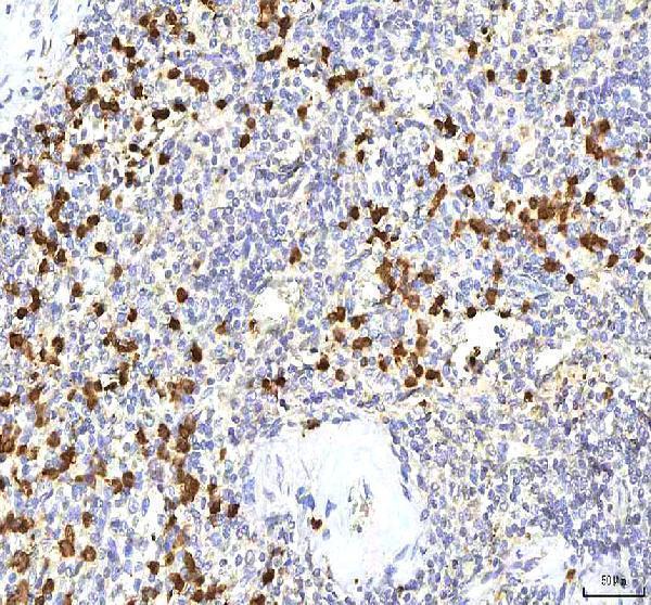
Figure 1. IHC analysis of S100A12 using anti-S100A12 antibody (A01478-1).
S100A12 was detected in a paraffin-embedded section of human spleen tissue. Heat mediated antigen retrieval was performed in EDTA buffer (pH 8.0, epitope retrieval solution). The tissue section was blocked with 10% goat serum. The tissue section was then incubated with 2 μg/ml rabbit anti-S100A12 Antibody (A01478-1) overnight at 4°C. Peroxidase Conjugated Goat Anti-rabbit IgG was used as secondary antibody and incubated for 30 minutes at 37°C. The tissue section was developed using HRP Conjugated Rabbit IgG Super Vision Assay Kit (Catalog # SV0002) with DAB as the chromogen.

Click image to see more details
Figure 2. IHC analysis of S100A12 using anti-S100A12 antibody (A01478-1).
S100A12 was detected in a paraffin-embedded section of human spleen tissue. Heat mediated antigen retrieval was performed in EDTA buffer (pH 8.0, epitope retrieval solution). The tissue section was blocked with 10% goat serum. The tissue section was then incubated with 2 μg/ml rabbit anti-S100A12 Antibody (A01478-1) overnight at 4°C. Peroxidase Conjugated Goat Anti-rabbit IgG was used as secondary antibody and incubated for 30 minutes at 37°C. The tissue section was developed using HRP Conjugated Rabbit IgG Super Vision Assay Kit (Catalog # SV0002) with DAB as the chromogen.

Click image to see more details
Figure 3. IHC analysis of S100A12 using anti-S100A12 antibody (A01478-1).
S100A12 was detected in a paraffin-embedded section of human spleen tissue. Heat mediated antigen retrieval was performed in EDTA buffer (pH 8.0, epitope retrieval solution). The tissue section was blocked with 10% goat serum. The tissue section was then incubated with 2 μg/ml rabbit anti-S100A12 Antibody (A01478-1) overnight at 4°C. Peroxidase Conjugated Goat Anti-rabbit IgG was used as secondary antibody and incubated for 30 minutes at 37°C. The tissue section was developed using HRP Conjugated Rabbit IgG Super Vision Assay Kit (Catalog # SV0002) with DAB as the chromogen.
Protein Target Info & Infographic
Gene/Protein Information For S100A12 (Source: Uniprot.org, NCBI)
Gene Name
S100A12
Full Name
Protein S100-A12
Weight
10.575kDa
Superfamily
S-100 family
Alternative Names
CAAF1; CAAF1Neutrophil S100 protein; CAAFI; CAGC; CAGCS100 calcium binding protein A12 (calgranulin C); CAGCS100; Calcium-binding protein in amniotic fluid 1; Calgranulin C; calgranulin-C; CGRPEN-RAGE; ENRAGE; EN-RAGE; Extracellular newly identified RAGE-binding protein; MRP6; p6calgranulin C; protein S100-A12; S100 calcium binding protein A12; S100 calcium-binding protein A12 (calgranulin C); S100 calcium-binding protein A12; S100A12 S100A12 CAAF1, CAGC, CGRP, ENRAGE, MRP-6, MRP6, p6 S100 calcium binding protein A12 protein S100-A12|EN-RAGE|calcitermin|calcium-binding protein in amniotic fluid 1|calgranulin C|extracellular newly identified RAGE-binding protein|migration inhibitory factor-related protein 6|neutrophil S100 protein
*If product is indicated to react with multiple species, protein info is based on the gene entry specified above in "Species".For more info on S100A12, check out the S100A12 Infographic

We have 30,000+ of these available, one for each gene! Check them out.
In this infographic, you will see the following information for S100A12: database IDs, superfamily, protein function, synonyms, molecular weight, chromosomal locations, tissues of expression, subcellular locations, post-translational modifications, and related diseases, research areas & pathways. If you want to see more information included, or would like to contribute to it and be acknowledged, please contact [email protected].
Specific Publications For Anti-S100A12 Antibody Picoband™ (A01478-1)
Hello CJ!
No publications found for A01478-1
*Do you have publications using this product? Share with us and receive a reward. Ask us for more details.
Recommended Resources
Here are featured tools and databases that you might find useful.
- Boster's Pathways Library
- Protein Databases
- Bioscience Research Protocol Resources
- Data Processing & Analysis Software
- Photo Editing Software
- Scientific Literature Resources
- Research Paper Management Tools
- Molecular Biology Software
- Primer Design Tools
- Bioinformatics Tools
- Phylogenetic Tree Analysis
Customer Reviews
Have you used Anti-S100A12 Antibody Picoband™?
Submit a review and receive an Amazon gift card.
- $30 for a review with an image
Be the first to review Anti-S100A12 Antibody Picoband™
*The first user to submit a review for a product is eligible for Boster's Innovating Scientists Reward, which gives product credits. This is in addition to the gift card reward.
Customer Q&As
Have a question?
Find answers in Q&As, reviews.
Can't find your answer?
Submit your question
